
On Dying of Dementia in a Capitalist System
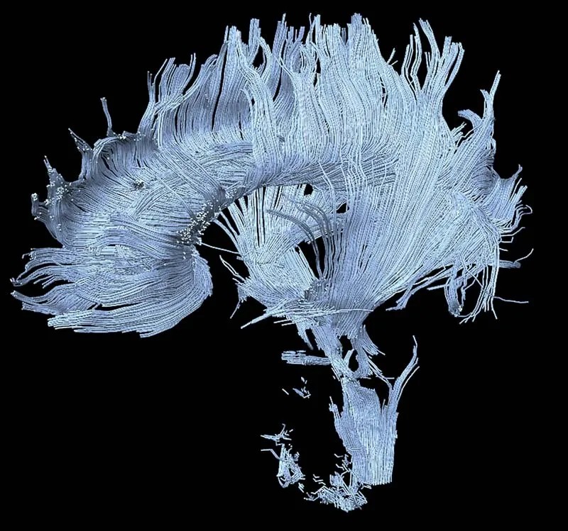
I find myself strangely disturbed by images of brain atrophy in various forms of dementia. I wonder if it is the implication of disorder and disease without cure, which shouldn’t be seen by a family member. Those MRI’s and Pet scans seem almost incomprehensible in spite of their clarity. So before progressing to atrophy, here is an image that can be viewed without disgust or despair, however fearsome the content.
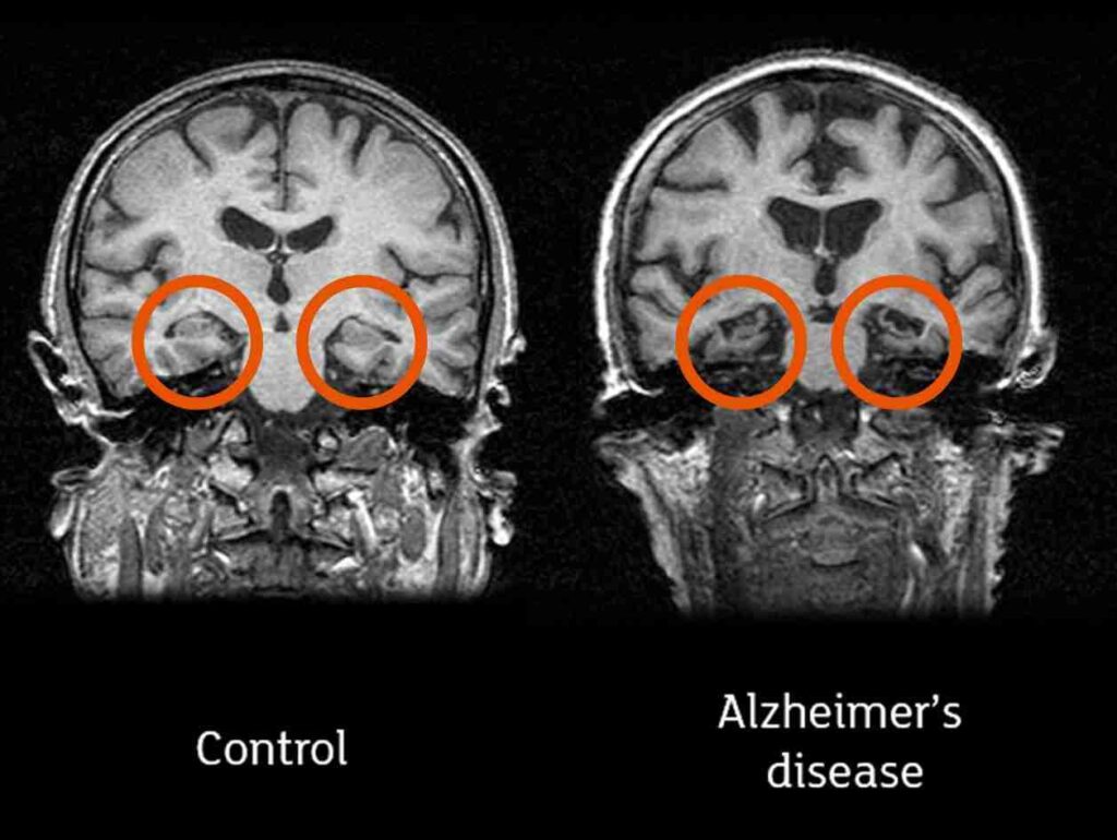
In Alzheimer’s disease, the hippocampi (circled in the image below) are often affected first. People with extensive damage are unable to form a/nd keep new memories. They are frequently disoriented.
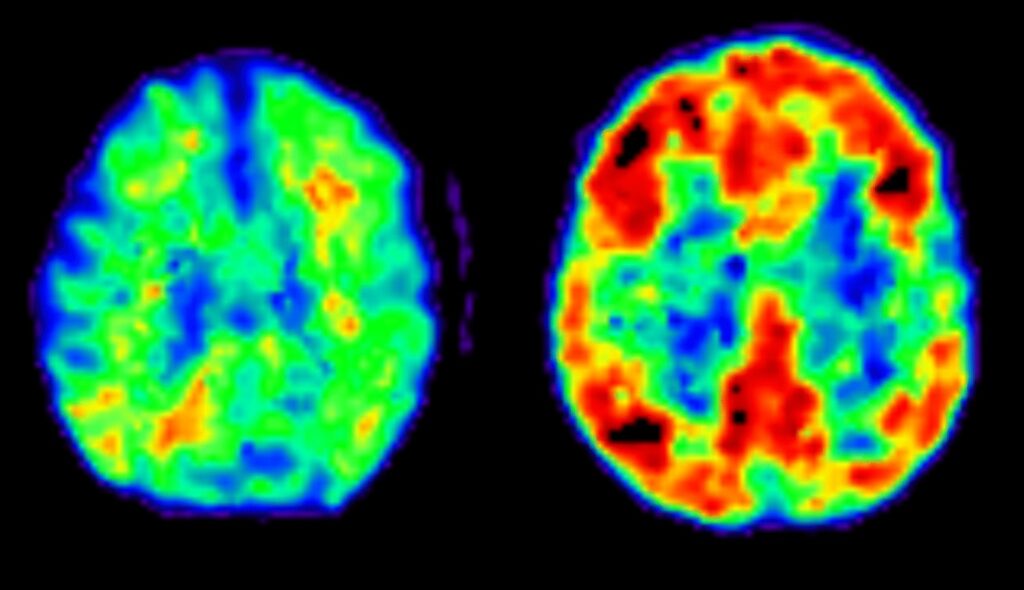
The images below are from a PET scan showing a protein called amyloid. Warmer colours (red) represent areas with more amyloid. The brain on the right is typical of someone with Alzheimer’s disease. There is evidence that clumps of the amyloid protein, tau, inflammation, and blood vessel disease cause brain cell death.
Different forms of dementia affect different parts of the brain resulting in different symptoms. This image compares a control to Alzheimer’s disease to Dementia with Lewy Bodies. Dementia with Lewy Bodies includes greater mood disturbance (depression, anxiety, apathy), visual hallucinations and delusions, fewer issues with memory at the start, and motor issues related to Parkinson’s disease.
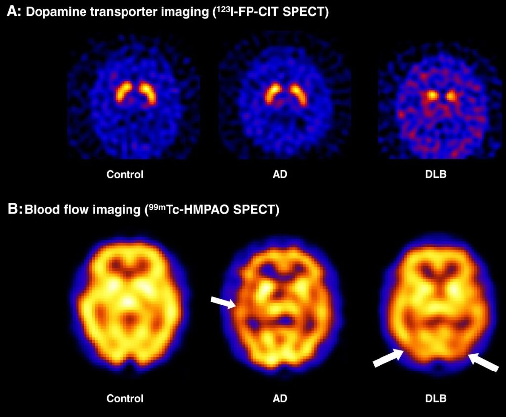
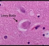
Lewy Body Dementia is associated with deposits of alpha-synuclein in the brain called Lewy Bodies.
This kind of distinction is important for 2 main reasons:
- Medication that is right for treatment in one form of dementia can produce negative impacts in another. Jim was given a medication for anxiety that is effective in Altzheimer’s disease but results in extreme confusion in Lewy Body Dementia. Anesthesia during a routine colonoscopy resulted in many hours of disorientation and hallucination.
- By the time symptoms appear, a great deal of damage to the brain has already occurred. The results of MRI and PET imaging can diagnose when treatment is more effective. This is called “prodromal.” Prodromal means before symptoms. (I had to look it up and now you won’t have to.)
Jim was not offered an MRI or a PET scan from the time we knew there was something wrong in 2014 until 2019 when he died. He did have an EEG which showed a “kind of slowing” of brain waves typical of both Alzheimer’s disease and Lewy Body disease. This test has re-emerged as a powerful diagnostic tool.
Additional forms of dementia include Vascular, Frontotemporal, HIV associated, Alcohol-related brain injury, and Chronic traumatic encephalopathy caused by repeated head injury.